The basic principles of three-dimensional (3D) ultrasound imaging mainly include three-dimensional geometric composition method, performance contour extraction method and voxel model method. The basic step of 3D ultrasonic imaging is to use a two-dimensional ultrasonic imaging probe to collect a series of 2D images in a certain spatial sequence and store them in the 3D reconstruction workstation. The computer performs spatial positioning on the 2D images collected according to a certain rule and compares the images. Image the gap between adjacent sections 2/12 The elements are supplemented and smoothed to form a 3D database, which is the post-processing of the image, and then the area of interest is delineated, 3D reconstruction is performed through the computer, and the reconstructed 3D image is displayed on the computer screen. 3D ultrasound imaging technology includes data acquisition, three-dimensional image reconstruction and three-dimensional image display. Baum and Greewood first proposed the concept of 3D ultrasound in 1961, but the development was relatively slow in the following 30 years. In the past ten years, with the continuous development of computer technology and ultrasound imaging technology, 3D ultrasound imaging technology has moved from the experimental research stage to the clinical application stage [2], which can be divided into (1) static 3D: collecting a certain number of 2D images and then making 3D group pictures, and then make various 3D displays, which are divided into 3D organ parenchyma and 3D blood vessel flow channels. (2)Dynamic
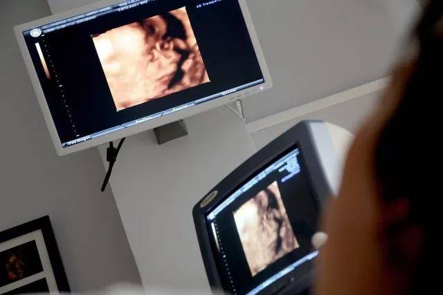
3D: Take multiple 2D images in different spaces at different time points and input and store them. Then use ECG to unify the time point, and combine the original images obtained at different times into a 3D image. The images will be assembled according to the ECG time series and then played back. Currently, it is widely used in various systems and parts such as heart, obstetrics and gynecology, small organs, blood vessels, and urogenital system [3]. Compared with 2D ultrasound, 3D ultrasound can display the three-dimensional anatomical shape and spatial relationship of tissue structures, has the advantages of intuitive image display, and can accurately measure medical diagnostic parameters.
Our contact number: +86 13027992113
Our email: 3512673782@qq.com
Our website: https://www.genosound.com/
Post time: Oct-27-2023